| 產品編號 | bsm-54462R |
| 英文名稱 | MARK3 Recombinant Rabbit mAb |
| 中文名稱 | 兔抗磷酸化絲氨酸/蘇氨酸蛋白激酶MARK3單克隆抗體 |
| 別 名 | C-TAK1; Cdc25C associated protein kinase 1; Cdc25C-associated protein kinase 1; CTAK1; ELKL motif kinase 2; EMK-2; Emk2; ETK 1; KIAA4230; KP78; MAP microtubule affinity regulating kinase 3; MAP/microtubule affinity-regulating kinase 3; Mark3; MARK3_HUMAN; |
| 研究領域 | 細胞生物 信號轉導 激酶和磷酸酶 |
| 抗體來源 | Rabbit |
| 克隆類型 | Recombinant |
| 克 隆 號 | 2F7 |
| 交叉反應 | Human,Mouse (predicted: Rat) |
| 產品應用 | IHC-P=1:100-500,IHC-F=1:200-400,IF=1:50-200,ICC/IF=1:50-500
not yet tested in other applications. optimal dilutions/concentrations should be determined by the end user. |
| 理論分子量 | 84 kDa |
| 檢測分子量 | |
| 細胞定位 | 細胞漿 細胞膜 |
| 性 狀 | Liquid |
| 濃 度 | 1mg/ml |
| 免 疫 原 | Recombinant human MARK3 protein: 600-750 |
| 亞 型 | IgG |
| 純化方法 | affinity purified by Protein A |
| 緩 沖 液 | 0.01M TBS (pH7.4) with 1% BSA, 0.02% Proclin300 and 50% Glycerol. |
| 保存條件 | Store at -20℃ for one year. Avoid repeated freeze/thaw cycles. The lyophilized antibody is stable at room temperature for at least one month and for greater than a year when kept at -20°C. When reconstituted in sterile pH 7.4 0.01M PBS or diluent of antib |
| 注意事項 | This product as supplied is intended for research use only, not for use in human, therapeutic or diagnostic applications. |
| PubMed | PubMed |
| 產品介紹 |
The protein encoded by this gene is activated by phosphorylation and in turn is involved in the phosphorylation of tau proteins MAP2 and MAP4. Several transcript variants encoding different isoforms have been found for this gene. [provided by RefSeq, Oct 2011] Function: Involved in the specific phosphorylation of microtubule-associated proteins for tau, MAP2 and MAP4. Phosphorylates CDC25C on 'Ser-216'. Regulates localization and activity of some histone deacetylases by mediating phosphorylation of HDAC7, promoting subsequent interaction between HDAC7 and 14-3-3 and export from the nucleus. Tissue Specificity: Ubiquitous. Similarity: Belongs to the protein kinase superfamily. CAMK Ser/Thr protein kinase family. MARK subfamily. Contains 1 KA1 (kinase-associated) domain. Contains 1 protein kinase domain. Contains 1 UBA domain SWISS: P27448 Gene ID: 4140 Database links: Entrez Gene: 4140 Human Entrez Gene: 17169 Mouse Omim: 602678 Human SwissProt: P27448 Human SwissProt: Q03141 Mouse Unigene: 35828 Human Unigene: 260504 Mouse Unigene: 408955 Mouse Unigene: 420865 Mouse Unigene: 23232 Rat |
| 產品圖片 |

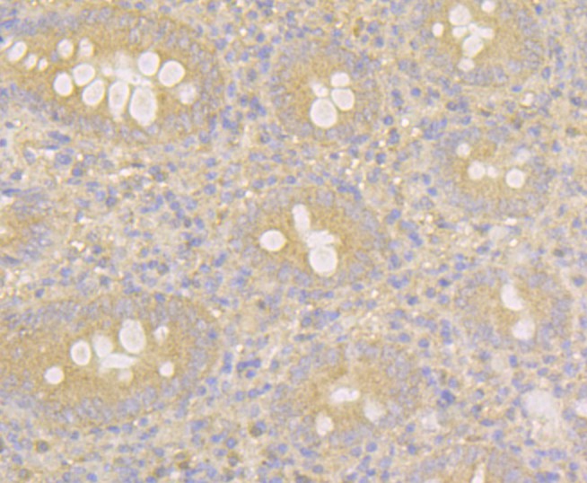
Immunohistochemical analysis of paraffin-embedded human breast cancer tissue using anti-MARK3 antibody. The section was pre-treated using heat mediated antigen retrieval with Tris-EDTA buffer (pH 8.0-8.4) for 20 minutes. The tissues were blocked in 5% BSA for 30 minutes at room temperature, washed with ddH2O and PBS, and then probed with the antibody (bsm-54462R) at 1/100 dilution, for 30 minutes at room temperature and detected using an HRP conjugated compact polymer system. DAB was used as the chrogen. Counter stained with hematoxylin and mounted with DPX.
Immunohistochemical analysis of paraffin-embedded human appendix tissue using anti-MARK3 antibody. The section was pre-treated using heat mediated antigen retrieval with Tris-EDTA buffer (pH 8.0-8.4) for 20 minutes. The tissues were blocked in 5% BSA for 30 minutes at room temperature, washed with ddH2O and PBS, and then probed with the antibody (bsm-54462R) at 1/100 dilution, for 30 minutes at room temperature and detected using an HRP conjugated compact polymer system. DAB was used as the chrogen. Counter stained with hematoxylin and mounted with DPX.

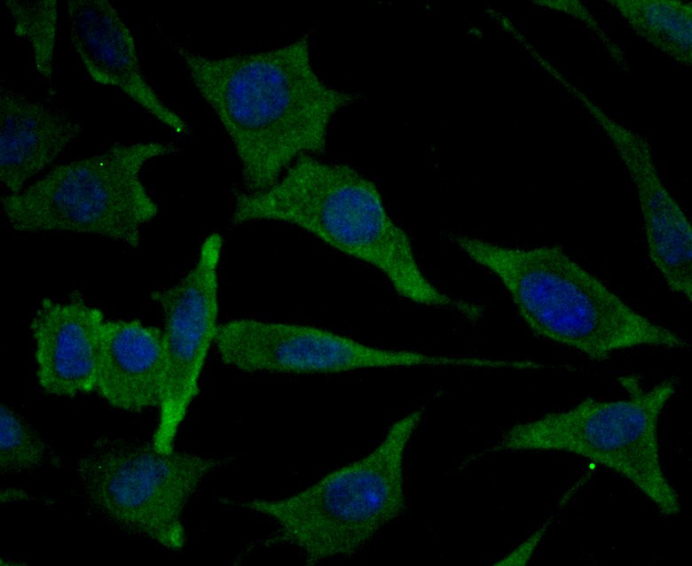
Immunocytochemistry staining MARK3 in SiHa cells (green). Formalin fixed cells were permeabilized with 0.1% Triton X-100 in TBS for 10 minutes at room temperature and blocked with 1% Blocker BSA for 15 minutes at room temperature. Cells were probed with MARK3 monoclonal antibody at a dilution of 1:50 for 1 hour at room temperature, washed with PBS. Alexa Fluorc? 488 Goat anti-Rabbit IgG was used as the secondary antibody at 1/100 dilution. The nuclear counter stain is DAPI (blue).
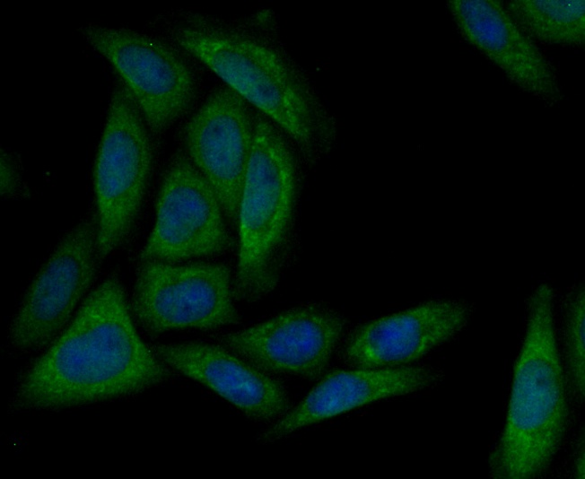
Immunocytochemistry staining MARK3 in SH-SY5Y cells (green). Formalin fixed cells were permeabilized with 0.1% Triton X-100 in TBS for 10 minutes at room temperature and blocked with 1% Blocker BSA for 15 minutes at room temperature. Cells were probed with MARK3 monoclonal antibody at a dilution of 1:50 for 1 hour at room temperature, washed with PBS. Alexa Fluorc? 488 Goat anti-Rabbit IgG was used as the secondary antibody at 1/100 dilution. The nuclear counter stain is DAPI (blue).
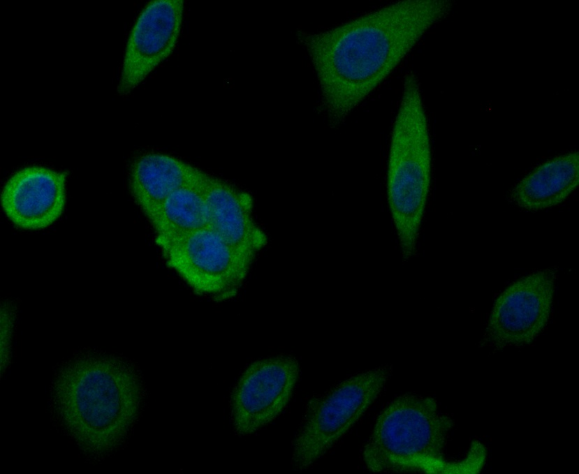
Immunocytochemistry staining MARK3 in MCF-7 cells (green). Formalin fixed cells were permeabilized with 0.1% Triton X-100 in TBS for 10 minutes at room temperature and blocked with 1% Blocker BSA for 15 minutes at room temperature. Cells were probed with MARK3 monoclonal antibody at a dilution of 1:100 for 1 hour at room temperature, washed with PBS. Alexa Fluorc? 488 Goat anti-Rabbit IgG was used as the secondary antibody at 1/100 dilution. The nuclear counter stain is DAPI (blue).

Immunohistochemical analysis of paraffin-embedded mouse small intestine tissue using anti-MARK3 antibody. The section was pre-treated using heat mediated antigen retrieval with Tris-EDTA buffer (pH 8.0-8.4) for 20 minutes. The tissues were blocked in 5% BSA for 30 minutes at room temperature, washed with ddH2O and PBS, and then probed with the antibody (bsm-54462R) at 1/100 dilution, for 30 minutes at room temperature and detected using an HRP conjugated compact polymer system. DAB was used as the chrogen. Counter stained with hematoxylin and mounted with DPX.

Immunohistochemical analysis of paraffin-embedded human appendix tissue using anti-MARK3 antibody. The section was pre-treated using heat mediated antigen retrieval with Tris-EDTA buffer (pH 8.0-8.4) for 20 minutes. The tissues were blocked in 5% BSA for 30 minutes at room temperature, washed with ddH2O and PBS, and then probed with the antibody (bsm-54462R) at 1/100 dilution, for 30 minutes at room temperature and detected using an HRP conjugated compact polymer system. DAB was used as the chrogen. Counter stained with hematoxylin and mounted with DPX.
Immunocytochemistry staining MARK3 in SiHa cells (green). Formalin fixed cells were permeabilized with 0.1% Triton X-100 in TBS for 10 minutes at room temperature and blocked with 1% Blocker BSA for 15 minutes at room temperature. Cells were probed with MARK3 monoclonal antibody at a dilution of 1:50 for 1 hour at room temperature, washed with PBS. Alexa Fluorc? 488 Goat anti-Rabbit IgG was used as the secondary antibody at 1/100 dilution. The nuclear counter stain is DAPI (blue).
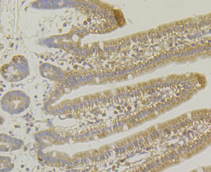
Immunohistochemical analysis of paraffin-embedded mouse small intestine tissue using anti-MARK3 antibody. The section was pre-treated using heat mediated antigen retrieval with Tris-EDTA buffer (pH 8.0-8.4) for 20 minutes. The tissues were blocked in 5% BSA for 30 minutes at room temperature, washed with ddH2O and PBS, and then probed with the antibody (bsm-54462R) at 1/100 dilution, for 30 minutes at room temperature and detected using an HRP conjugated compact polymer system. DAB was used as the chrogen. Counter stained with hematoxylin and mounted with DPX.
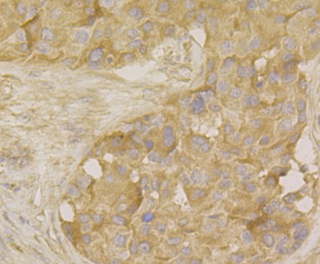
Immunohistochemical analysis of paraffin-embedded human breast cancer tissue using anti-MARK3 antibody. The section was pre-treated using heat mediated antigen retrieval with Tris-EDTA buffer (pH 8.0-8.4) for 20 minutes. The tissues were blocked in 5% BSA for 30 minutes at room temperature, washed with ddH2O and PBS, and then probed with the antibody (bsm-54462R) at 1/100 dilution, for 30 minutes at room temperature and detected using an HRP conjugated compact polymer system. DAB was used as the chrogen. Counter stained with hematoxylin and mounted with DPX.

Immunocytochemistry staining MARK3 in SH-SY5Y cells (green). Formalin fixed cells were permeabilized with 0.1% Triton X-100 in TBS for 10 minutes at room temperature and blocked with 1% Blocker BSA for 15 minutes at room temperature. Cells were probed with MARK3 monoclonal antibody at a dilution of 1:50 for 1 hour at room temperature, washed with PBS. Alexa Fluorc? 488 Goat anti-Rabbit IgG was used as the secondary antibody at 1/100 dilution. The nuclear counter stain is DAPI (blue).

Immunocytochemistry staining MARK3 in MCF-7 cells (green). Formalin fixed cells were permeabilized with 0.1% Triton X-100 in TBS for 10 minutes at room temperature and blocked with 1% Blocker BSA for 15 minutes at room temperature. Cells were probed with MARK3 monoclonal antibody at a dilution of 1:100 for 1 hour at room temperature, washed with PBS. Alexa Fluorc? 488 Goat anti-Rabbit IgG was used as the secondary antibody at 1/100 dilution. The nuclear counter stain is DAPI (blue).
|
| 1、抗體溶解方法 | |
| 2、抗體修復方式 | |
| 3、常用試劑的配制 | |
| 4、免疫組化操作步驟 | |
| 5、免疫組化問題解答 | |
| 6、Western Blotting 操作步驟 | |
| 7、Western Blotting 問題解答 | |
| 8、關于肽鏈的設計 | |
| 9、多肽的溶解與保存 | |
| 10、酶標抗體效價測定程序 | |